Stress Echocardiogram
What is a Stress Echocardiogram
Exercise stress echocardiography, which is usually shortened to exercise stress echo or ESE, is a test combining exercise treadmill testing and cardiac ultrasound (echocardiography) to diagnose abnormalities of the cardiac chambers and coronary arteries.
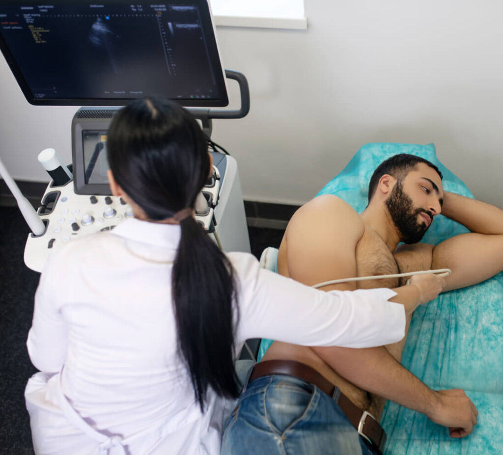
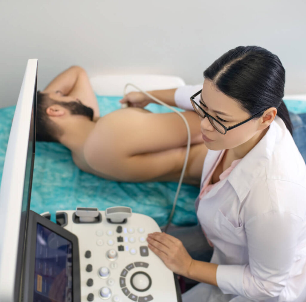
This test uses ultrasound pictures of the heart (also called echocardiography, or echo) to assess the state of the pumping chamber of the heart (the left ventricle) at rest. The patient then exercises on a treadmill, to speed their heart rate up. This increases the demands on the heart. The blood supply to the heart, via the coronary arteries, has to increase to meet these demands. After the treadmill is stopped, further images of the pumping chamber of the heart are recorded. This allows the pumping action of the heart to be assessed after exercise, compared to at rest.
Because exercise stress echo uses heart ultrasound as well as ECG, it is usually more accurate than treadmill testing because of the additional information collected. In addition, it does not involve needles or exposure to medical radiation.
Principles of Stress Echocardiogram
The pumping chamber of the heart responds to exercise by an increase in heart rate and an increase in contractility (the pumping action of the heart). Blood pressure also increases with exercise. If there is a blocked coronary artery, the part of heart muscle past the blockage does not obtain sufficient blood supply when exercise is performed.
This is because when the heart exercises, it requires a higher blood flow to each part of the heart. This increased blood supply to the muscular wall of the heart occurs by increased blood flow in the coronary arteries. If one or several coronary arteries are blocked, a normal increase in pumping action is not seen. One part of the heart muscle will not increase contractility normally.
What sort of patients undergo this test?
There are three main groups of patients who undergo stress echo.
The first group are those who have experienced cardiac symptoms – mainly chest discomfort or shortness-of-breath. In these patients, the referring doctor wants to know if coronary heart disease (narrowing of the coronary arteries) could be causing these symptoms.
The second group are patients who have had a exercise stress test without ultrasound imaging, and have an abnormal resting ECG. These patients require ultrasound imaging so the stress test can be analysed. Other patients may have ECG changes during stress testing that are a technical anomaly rather than reflecting the presence of heart disease. Stress echocardiography can very often diagnose or exclude heart disease in these patients.
The third group of patients are those who have established heart disease: for example, a previous heart attack (myocardial infarction), and those whom have intra-coronary stents or whom have had coronary artery bypass grafting (cardiac bypass surgery). In these patients, cardiologists use stress echo to ensure the contractility of the heart chambers after exercise is normal, which suggests the blood flow through the stents and bypass grafts is normal. Stress echo is a good way of monitoring patients’ progress over time, as it does not involve medical radiation, and it allows direct imaging with ultrasound of the chambers of the heart.
How should I prepare for a Stress Echocardiogram?
Please wear comfortable walking or running shoes. Women should wear a top and pants or shorts, rather than a dress. Do not eat a heavy meal prior to the test. However, you do not need to fast before this test. You will be asked to withhold any tablets that slow down the heart rate including beta-blockers (such as Metoprolol, Diltiazem and Isoptin). You will be asked about what medication you are on at the time of booking and will be advised on when to stop these medications prior to the exercise stress echo.
If stopping these tablets causes problems for you, such as high blood pressures or palpitations, please discuss this with the staff at the time of booking, as it may be possible for you to still do the test while on your normal medication.
What is involved?
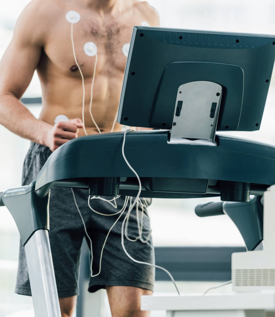
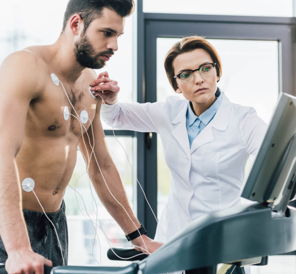
You will be asked to put a gown on. Small sticky ECG dots will be applied to the front and left side of your chest to record an ECG during the test, and a blood pressure cuff will be applied to your arm. You will lie down on a couch so the cardiologist can record the ultrasound images of your heart at rest.
You will then walk on a treadmill for a few minutes until your heart rate has increased. Your blood pressure and a continuous ECG (heart rhythm trace) will be recorded, and the nurse and doctor will ask you if you feel symptoms such as chest pain. The treadmill will stop when your heart rate is adequate for the test (usually between 110 and 150 beats per minute, depending on your age). You will lie back down on the couch, and the images of your rapidly beating heart (called the post exercise images) will be recorded. Your ECG and blood pressure will be continued to be monitored in this recovery phase. The cardiologist will compare the resting images of your heart to the images taken after exercise and decide whether all areas of the heart muscle increase contractility normally in response to exercise.
Most people do not need to jog or run on the treadmill to increase their heart rate to the required amount. Usually, a brisk walk is sufficient.We use a standardised treadmill protocol which is used in most countries of the world for stress testing, called the Bruce protocol. This treadmill protocol starts with a slow 3-minute warm up phase, before the treadmill gradient and speed increases. The pace and gradient increase a little every three minutes until the desired heart rate is reached. Most patients walk for between 4 to 8 minutes on the treadmill. Some patients can complete as much as 12 minutes.
If a patient wants to stop for any reason before the test is completed, the test can be interrupted and the peak heart images can still be collected.
How long will it take?
Although most patients walk on the treadmill for 4 to 8 minutes, we give a longer booking time for this test (45-60 minutes) to allow time for you and for the ultrasound machine to be set-up, to allow plenty of time for recovery, to allow completion of the recording of all the information needed, and for preparation of the report of your test, which is returned to your referring doctor.
What happens next?
If your test is normal, it is likely you will not need any further cardiac tests.
If the test shows an abnormality with the pumping chamber of the heart after exercise, you may need a coronary angiogram, which is a dye injection test of the coronary arteries. If you need this test, it can be organised for you at the same appointment.
The report will be sent to your referring doctor on the day you have the test.
What if I cannot walk easily?
Many patients cannot walk easily due to musculoskeletal problems of the knees, hips, ankles or back. If you can walk, but find a flat gradient or a slower speed easier, you may still be able to use the treadmill very successfully. We have a range of protocols available, some with a flatter gradient and slower treadmill speed.
If you are severely disabled and cannot walk at all, stress echo can still be performed, but not using the treadmill. In this case we use a short intravenous infusion of a cardiac stimulant medication, called Dobutamine. This acts on the heart directly to increase the pumping action of the heart and the heart rate. This medication is infused gradually to increase the heart rate to the desired level. The patient spends the entire test lying down on the echo couch.
How do I get more information?
Our clerical staff can answer your questions at the time of booking. The supervising nurse and doctor will also be happy to discuss any questions or concerns you may have.